(Click here for self-help products that can help with ADD and other learning concerns.)
David Noton, PhD
The Forest Institute
Reprinted from J. of Neurotherapy, 2(2):8-13, 1997.
ABSTRACT
Two studies of premenstrual syndrome (PMS), EEG, and photic stimulation
have recently been completed at the Royal Postgraduate Medical School,
Hammersmith Hospital, London (UK). In a preliminary trial of photic
stimulation as a treatment for PMS seventeen women with PMS were treated
with a take-home flashing light device for 15 to 20 minutes per day
throughout their cycle. At the end of three months of treatment the
median reduction in PMS symptoms for the 17 patients was 76% and twelve
of the 17 patients technically no longer had PMS. Separately, an EEG
study of six women with PMS demonstrated that, when they were
premenstrual, their EEGs showed more slow (delta) activity and slower
P300 evoked response than when they were mid-cycle. These results are
discussed in the context of other known “slow brainwave”
disorders, such as ADD and Minor Head Injury, and various theoretical
explanations are proposed.
EXPERIMENTAL RESULTS
Preliminary Trial of Photic Stimulation for PMS
A preliminary trial of photic stimulation (flashing light therapy) as a
treatment for PMS was recently completed by Duncan Anderson and his
associates at the Royal Postgraduate Medical School, Hammersmith
Hospital, London (UK). It was an open study of 17 women, all of whom had
confirmed, severe, and long-standing PMS.
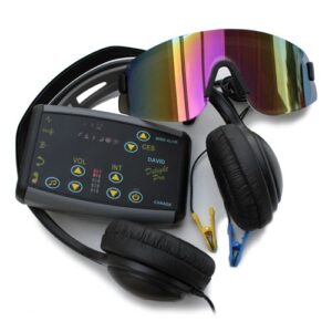
The flashing light device is similar to the device previously used for
treatment of migraine (Anderson 1989). It consists of a mask, which
covers the eyes shutting out all light. Mounted in the mask are red LED
lamps, one over each eye, which flash alternately in left and right
eyes. The device is portable and designed to be used by the patient at
home. The brightness of the light and the frequency of flashing are
controlled by the patient, with ranges of approximately 10 to 45 mcd and
0.5 to 50 Hz respectively (one frequency cycle consisting of light in
the left eye for half the cycle and then light in the right eye for half
the cycle). The patients were instructed to start at the brightest
setting and at the flicker-fusion point (around 30 Hz) and then adjust
the brightness and frequency for best comfort. The patients were asked
to use the device for 15 minutes per day, every day throughout their
menstrual cycle. The patients recorded their symptoms daily for two
menstrual cycles before treatment, three cycles during treatment, and
one cycle after treatment was stopped.
At the end of treatment the median reduction in PMS symptoms for the 17
patients was 76%. Twelve of the 17 patients technically no longer had
PMS. Although the results of an open trial are subject to placebo
effects, the results were so large and persistent that it is unlikely
that placebo can fully explain them. The complete results of this trial
are published in detail elsewhere (Anderson et al. 1997).
Study of EEG during PMS
A study of EEG during PMS was recently completed by Istra Toner and her
associates at the Royal Postgraduate Medical School in London (Toner et
al. 1995). Six women with self-reported PMS had 21 channel QEEG
recordings and P300 evoked potentials measured during mid-cycle and
premenstrually. Ages ranged from 30 to 43 and all were taking no
treatment for PMS.
A significant increase in delta activity during PMS was observed
(p=0.043) along with a suggestive, but not necessarily significant,
decrease in beta activity. This is consistent with previous reports of
increased slow activity and decreased fast activity during PMS (Harding
et al. 1976, Lamb et al. 1953).
P300 evoked potential was elicited using an odd-tone procedure with a
frequent tone (1000 Hz) and an odd tone (2000 Hz) presented in the ratio
4:1 at a rate of 1 per second. Using global field power averaging, a
significant increase in P300 latency during PMS was observed (p=0.027).
DISCUSSION AND INTERPRETATION
PMS is a “Slow Brainwave” Disorder
It is proposed that there is a group of disorders characterized by
excessive low frequency EEG activity. For example:
| Disorder | Abbr. | Reported Brainwave Characteristic | Attention Deficit | ADHD | Excess theta/beta ratio (Lubar 1991, etc.) | Chronic Fatigue Syndr | CFS | Slow alpha, excess theta (Lindenfeld et al 1996) | Minor Head Injury | MHI | Diffuse slow activity (Duffy et al. 1989, Ayers 1987, both also quoted in Byers, 1995) |
Toxic Trauma | TT | Excess slow activity (Heuser 1994) | Premenstrual Syndrome | PMS | Excess delta, slow P300 (Toner 1995reported above) |
Based on the EEG results described above, PMS is seen to belong to this
group of disorders.
Treatment of Slow Brainwave Disorders with Photic Stimulation
The preliminary trial reported above shows the efficacy of photic
stimulation as a treatment for PMS. The treatment of ADHD with photic
stimulation has been developed extensively by Harold Russell and his
associates, using frequencies of 18 Hz and 10 Hz alternating for two
minute periods, with demonstrable improvements in IQ scores and behavior
(Russell and Carter, 1993). Many clinicians appear to be using photic
stimulation informally for ADHD and the other slow brainwave disorders,
with anecdotal reports of successful treatment but with very few
published results.
Treatment of Slow Brainwave Disorders with Neurofeedback
Many neurofeedback (EEG biofeedback) practitioners report successful
treatment of some or all of these slow brainwave disorders. For example,
the Lubar’s have for many years worked with children with ADHD, training
them with beta frequency biofeedback, with excellent results (Lubar 1991
and 1989); the Othmer’s have a long history of success with beta
frequency biofeedback with patients with all of the disorders in this
group (Othmer 1994); and there are many other practitioners using this
approach. Generally the feedback protocol involves positive
reinforcement of beta frequencies and negative reinforcement of theta
frequencies, though various other protocols are also used successfully.
The Brainwave Frequency Hypothesis
A reasonable explanation that is commonly proposed for the above
experimental and clinical results is that the key to treating these
disorders (all characterized by excessive slow brainwave activity) is to
speed up the brainwave frequency. It is proposed that this can be
accomplished either by training the patients to speed up their own
brainwaves (beta-training neurofeedback) or by entraining the patients’
brainwaves with a photic stimulation device flashing at beta
frequencies.
Problems with the Brainwave Frequency Hypothesis
Unfortunately there is evidence, both from photic stimulation research
and from neurofeedback training, that undermines this brainwave
frequency hypothesis.
In the trial of PMS and photic stimulation reported above, the patients
were free to adjust the frequency of the flashing light at will, between
0.5 Hz and 50 Hz. A frequency of around 30 Hz (high beta) was suggested,
based on previous clinical results, but the patients were free to change
this at any time in any session. Of those patients who achieved a
greater than 50% reduction in symptoms, about half chose to operate the
flashing light in the range of 5 to 10 Hz, ie, theta-alpha frequency,
not beta frequency.
Furthermore, some neurofeedback clinicians report equally good results
when treating slow brainwave disorders with frequency protocols quite
different from the beta enhancement/theta reduction protocol discussed
above. In fact, Hoffman et al. (1995) list six different neurofeedback
protocols (including alpha training) that have been used successfully
for minor head injury.
Apparently “speeding up” the brainwaves with photic
stimulation or neurofeedback at beta frequencies is not an adequate
explanation for the successful treatment of these disorders.
The Cerebral Blood Flow Hypothesis
Many studies have shown that excessive slow brainwave activity is
closely associated with hypoperfusion, ie, insufficient cerebral blood
flow. These studies have been collected and summarized by Toomim (1994).
Looking at the individual “slow brainwave” disorders we see
that in each case there is some evidence for hypoperfusion:
| Disorder | Evidence for Insufficient Cerebral Blood Flow | ADHD | Localised hypoperfusion demonstrated by Zametkin et al. (1990) | CFS | Hypoperfusion caused by hypotension, Bou-Holaigah et al. (1995) |
MHI | Hypoperfusion demonstrated by Ichise et al. (1994) | TT | Localised hypoperfusion demonstrated by Heuser et al. (1994) | PMS | Preliminary SPECT tests show localised cerebral hypoperfusion (Amen, 1996) |
The causal relationship between slow brainwave activity and
hypoperfusion is unclear. It is possible that reduced neuronal activity
demands less blood flow or that reduced blood flow causes reduced
neuronal activity or even that there is a “vicious circle”
with neither component being able to initiate recovery.
However, it is known that cerebral blood flow is increased by photic
stimulation (for example Sappey-Marinier et al., 1992 and Fox et al.,
1988). It is possible that this is the mechanism by which photic
stimulation relieves PMS and other slow brainwave disorders.
The Role of Frequency
This is not to suggest that frequency is without significance. The
training frequency in neurofeedback and the flash frequency in photic
stimulation have been shown to encourage or entrain brainwaves of that
frequency and this may have therapeutic value independent of blood flow
considerations, by training the patient’s brainwaves to operate at
beneficial frequencies. And in some cases, for example alpha-theta
neurofeedback, frequency is obviously critical, enabling the patient to
access early emotional material of great therapeutic importance.
However, Othmer (1996) has suggested that a major component of
neurofeedback training is the exercising and training of the mechanisms
of arousal and attention, regardless of the frequency which is being
trained. Exercising these mechanisms might be expected to result in an
increase in neuronal activity and associated cerebral blood flow.
SUMMARY AND CONCLUSIONS
PMS and EEG
An EEG study of six women with PMS demonstrated that, when they were
premenstrual, their EEGs showed more slow (delta) activity and slower
P300 evoked response than when they were mid-cycle. It is concluded that
PMS belongs to a group of disorders characterized by excessive slow
brainwave activity.
PMS and Photic Stimulation
In a preliminary trial of photic stimulation as a treatment for PMS
seventeen women with PMS treated themselves with a take-home flashing
light device for 15 to 20 minutes per day throughout their cycle.
Thirteen of the seventeen experienced a greater than 50% reduction in
their symptoms. It is concluded that photic stimulation is an effective
treatment for PMS.
Brainwave Frequency vs. Cerebral Blood Flow
Some of the other “slow brainwave” disorders are also being
treated effectively with photic stimulation and all of the disorders are
being successfully treated with beta frequency neurofeedback. This has
led to the common hypothesis that these treatments are effective because
they “speed up” the brainwaves, but in fact, at least with
these “slow brainwave” disorders, the frequency used in the
treatment, whether photic stimulation or neurofeedback, seems to be of
secondary importance. It is suggested that increases in cerebral blood
flow and associated increases in neuronal activity may be of equal or
greater significance.
Photic Stimulation vs. Neurofeedback
If both neurofeedback and photic stimulation are effective in the
treatment of these “slow brainwave” disorders, perhaps the
best treatment may often be a combination of the two. Photic stimulation
has the advantages of low cost and portability; it can be given to
patients as “homework” between sessions and as pre-training
for neurofeedback, to “teach” the brain the frequency that is
to be trained. Neurofeedback develops the patient’s sense of
self-control and also has the unique advantage of localisation, the
ability to affect neuronal activity and brain blood flow specifically at
a training site chosen for its relevance to the disorder, rather than
just in the cortex in general. The combination of neurofeedback and
photic stimulation seems particularly appropriate for ADHD, where the
patient may initially have motivational difficulties with the
neurofeedback training and need assistance from any other modality
available.
REFERENCES
Amen, D., Personal communication, 1996.
Anderson, D.J., “The Treatment of Migraine with Variable Frequency
Photo-stimulation,” Headache, 29:154-155, 1989.
Anderson, D.J, Legg, N.J., Ridout, D.A., “Preliminary trial of
photic stimulation for premenstrual syndrome,” J. of Obstetrics and
Gynaecology, 17(1):76-79, 1997.
Ayers, M.E., “Electro-encephalographic neurofeedback and closed
head injury of 250 individuals,” A paper presented at the National
Head Injury Foundation Annual Conference, 1987 (quoted in Byers (1995)).
Bou-Holaigah, I., Rowe, P.C., Kan, J., Calkins, H., “The
Relationship Between Neurally Mediated Hypotension and the Chronic
Fatigue Syndrome,” JAMA, 274:961-967, 1995.
Byers, A.P., “Neurofeedback Therapy for a Mild Head Injury,”
J. of Neurotherapy, 1(1):22-37, 1995.
Duffy, F.H., Iyer, V.G., Surwillo, W.W., Clinical electroencephalography
and topographic brain mapping: Technology and practice, Springer-Verlag,
New York, Berlin, 1989 (quoted in Byers (1995)).
Fox, P.T., Raichle, M.E., Mintum, M.A., Dence, C., “Nonoxidative
glucose consumption during focal physiologic neural activity,”
Science, 241:462-464, 1988.
Harding, G. F. A., Thompson, C. R. S., “EEG Rhythms and internal
milieu.” In: Remond, A. Handbook of Electroencephalography and
Clinical Neurophysiology (Vol 6A, pp. 176-194), 1976. Amsterdam:
Elsevier.
Heuser, G., Mena, I., Alamos, F., “NeuroSPECT Findings in Patients
Exposed to Neurotoxic Chemicals,” Toxicology and Industrial Health,
10(4/5): 561-571, 1994.
Hoffman, D.A., Stockdale, S., Hicks, L.L., Schwaninger, J.E.,
“Diagnosis and Treatment of Head Injury,” J. of Neurotherapy,
1(1):14-21, 1995.
Ichise, M., Chung, D., Wang, P., Wortzman, G., Gray, B., Franks, W.,
“Technetium-99-HMPAO SPECT, CT and MRI in the evaluation of
patients with chronic traumatic brain injury: a correlation with
neuropsychological performance,” J. of Nuclear Medicine,
35(2):217-225, 1994.
Lamb, W., Ulett, G., Masters, W., Robinson, D., “Premenstrual
tension EEG, hormonal and psychiatric evaluation.” American J.
Psychiatry, 109: 840-848, 1953.
Lindenfeld, K.M., Budzynski, T., Andrasik, F., “EEG Patterns and
Chronic Fatigue Syndrome,” (Abstract) Proc. AAPB 27th Annual
Meeting, Albuquerque, NM, 1996.
[A more complete report, not available at the time this paper was being
prepared, is:
Billiot, K. M., Budzynski, T.H., Andrasik, F., “EEG Patterns and
Chronic Fatigue Syndrome,” J. of Neurotherapy, 2(2):20-30, 1997.]
Lubar, J.F., “Discourse on the development of EEG diagnostics and
biofeedback for attention-deficit/hyperactivity disorders,”
Biofeedback and Self-Regulation, 16(3):201-225, 1991.
Lubar, J. F., “Electroencephalographic biofeedback and neurological
applications.” In J. V. Basmajian (Ed.), Biofeedback Principles and
Practice for Clinicians (3rd ed.), pp.67-90, 1989. Baltimore: Williams
& Wilkins.
Othmer, S.O., personal communication, 1996.
Othmer, S.O., “EEG Biofeedback Training,” Megabrain Report, J.
of Mind Technology, 2(3):43-47, 1994.
Russell, H.L., and Carter, J.L., “A Pilot Investigation of Auditory
and Visual Entrainment of Brainwave Activity in Learning-Disabled
Boys,” Texas Researcher, J. of the Texas Center for Educational
Research, 4:65, 1993.
Sappey-Marinier, D., Calabrese, G. , Fein, G., Hugg, J.W., Biggins, C.,
Weiner, M.W., “Effect of Photic Stimulation on Human Visual Cortex
Lactate and Phosphates using 1H and 31P Magnetic Resonance
Spectroscopy,” J. of Cerebral Blood Flow and Metabolism,
12:584-592, 1992.
Toner, I., Peden, C., Carol, S., Hayden, M., Stone, J., Vucicevic, V.,,
“P300 and QEEG changes during menstrual cycle.” (Abstract)
International Journal of Psychophysiology, 1995.
Toomim, H., “Brain Blood Flow and Neurofeedback,” Biocomp
Research Institute, Culver City, CA, 1994.
Zametkin, A.J., Nordahl, T.E., Gross, M., King, A.C., Semple, W.E.,
Rumsey, J., Hamburger, S., Cohen, R.M., “Cerebral Glucose
Metabolism in Adults with Hyperactivity of Childhood Onset,” The
New England Journal of Medicine, 323(20):1361-1366, 1990.